Thorax Anatomy Illustration High Biology Diagrams The thorax is the superior part of the trunk extending between the neck and the abdomen.It consists of several components:. Thoracic wall; Several cavities; Nerves, blood vessels, lymphatics; Internal organs; Breasts; Thoracic wall. The thoracic wall consists mainly of muscles and bones that form the thoracic cage.Overall, the thoracic wall is formed by the following structures:
Anatomy of the Thorax Shari L. Meyerson David A. Harpole Jr. The bony and soft tissue components of the chest wall combine to create an anatomic space, which houses some of the most vital structures in the human body. The thoracic skeleton creates a protected space for the heart. It provides a protective framework for…

Thorax: Anatomy, wall, cavity, organs & neurovasculature Biology Diagrams
Anatomy. The thorax, commonly known as the chest, is a vital region of the human body that provides protection to critical organs, Sobotta Atlas of Human Anatomy: Volume 2 - Thorax, Abdomen, and Pelvis (14th ed.). Urban & Fischer. ISBN 978-0723434521. Ellis, H. (2006). The thoracic wall consists of a bony framework held together by twelve thoracic vertebrae posteriorly, giving rise to ribs that encircle the lateral and anterior thoracic cavity. The first nine ribs curve around the lateral thoracic wall and connect to the manubrium and sternum. Ribs 10 to 12 are relatively short and attach to the costal margins of the ribs just above them. Ribs 10 to 12, due Your thoracic cavity is the large space in your chest where some of your body's most important work gets done. If you're interested in learning about the thoracic cavity, you may have had a recent lung or heart disease diagnosis. Or maybe you just want to know more about the human body. Either way, your healthcare provider can help you
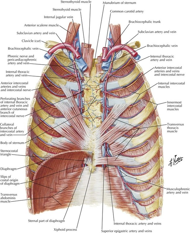
The organs of the thorax include the thymus gland, the breasts, the heart, the lungs, the tracheobronchial tree and the pleurae. The thymus gland is located in the superior mediastinum of the thoracic cavity but may also extend into the neck.It is classified as a lymphoid organ, meaning that it plays a role in the development of the immune system. Thoracic wall The first step in understanding thorax anatomy is to find out its boundaries. The thoracic, or chest wall, consists of a skeletal framework, fascia, muscles, and neurovasculature - all connected together to form a strong and protective yet flexible cage.. The thorax has two major openings: the superior thoracic aperture found superiorly and the inferior thoracic aperture The thorax (pl.: thoraces or thoraxes) [1] or chest is a part of the anatomy of mammals and other tetrapod animals located between the neck and the abdomen. [2] [3]In insects, crustaceans, and the extinct trilobites, the thorax is one of the three main divisions of the body, each in turn composed of multiple segments.. The human thorax includes the thoracic cavity and the thoracic wall.

Thorax (overview) Biology Diagrams
The thorax forms from the thoracic wall, its superficial structures (breast, muscles, and skin), and the thoracic cavity. A thorough comprehension of the anatomy and function of the thorax will help identify, differentiate, and treat the plethora of pathology that can occur within the thorax. The thorax is the area of the body situated between the neck and the abdomen. The thorax itself can be split up into various areas that contain important structures.. The thorax is bound by bony structures including the 12 pairs of ribs and thoracic vertebrae, whilst also being supported by many ligaments and muscles.. The muscles of the thorax are also important for the vital actions of