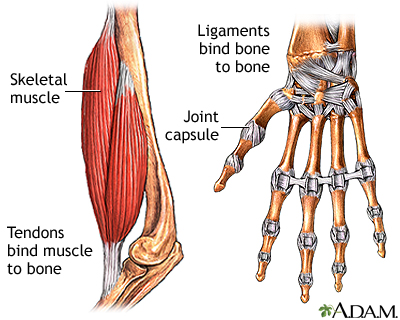
Tendinitis Biology Diagrams Achilles tendon: This runs from the calf muscle to the heel.It is the strongest and largest tendon in the body, making it possible to run, jump, climb stairs, and stand on your toes. Tibialis posterior: This attaches the calf muscle to the bones on the inside of the foot and supports the arch of the foot.; Tibialis anterior: This runs from the outer bone of the lower leg to the tarsals and Because they are so complicated, human feet can be especially prone to injury. Strains, sprains, tendonitis, torn ligaments, broken bones, fallen arches, bunions, corns, and plantar warts can all occur. Here we will talk more about the anatomy of the human foot and its many moving parts.
What is the anatomy of a tendon? Tendons are mostly collagen, one of the most abundant proteins in your body. Tendons also contain blood vessels and nerves. Advertisement. Collagen fibers are flexible, strong and resistant to damage. A tendon's structure is similar to a fiberoptic cable or a rope, with small collagen fibers arranged in bundles.

Leg Anatomy: Complete Guide with Parts, Names & Diagram Biology Diagrams
Tendons are dense connective tissue structures, composed of an hierarchy of longitudinally arranged collagen fibers, elastin, glycoproteins, proteoglycans and a lesser amount of specialized fibroblast cells.. Type 1 collagen is the most abundant form of collagen identified within tendinous structures and is directly responsible for its strength and durability.
The tendon is a "mechanical bridge," transmitting muscle forces to the bones and joints. This tough, fibrous structure also helps muscles complete joint movements along a plane. The tendon type reflects its associated muscle's morphology and function. Tendon tissue is present throughout an entire muscle's length, not only the tips. The muscle's connective tissue layers (epimysium, perimysium Its tendon begins slightly above the outside ankle bone, wraps around the ankle, and attaches to the outside edge of the fifth toe bone. Leg Anatomy: Joint Human Head. Skull Anatomy: Complete Guide with Parts, Names, Functions & Diagram; Ultimate Guide to Eye Anatomy: Parts, Structure, Functions & Diagram A solid understanding of anatomy is essential to effectively diagnose and treat patients with foot and ankle problems. Anatomy is a road map. Most structures in the foot are fairly superficial and can be easily palpated. Anatomical structures (tendons, bones, joints, etc) tend to hurt exactly where they are injured or inflamed.
:max_bytes(150000):strip_icc()/tendonitis-definition-causes-treatment-2696478-v21-5c77259146e0fb0001a9830f.png)
Symptoms and causes Biology Diagrams
Tendons are situated between bone and muscles and are bright white in colour, their fibro-elastic composition gives them the strength require to transmit large mechanical forces. Each muscle has two tendons, one proximally and one distally. The point at which the tendon forms attachment to the muscle is also known as the myotendinous junction (MTJ) and the point at which it attaches to the
