Placenta and Placental Development Biology Diagrams The placenta is a unique vascular organ that receives blood supplies from both the maternal and the fetal systems and thus has two separate circulatory systems for blood: (1) the maternal-placental (uteroplacental) blood circulation, and (2) the fetal-placental (fetoplacental) blood circulation. The uteroplacental circulation starts with the maternal blood flow into the intervillous space The placenta is arguably the most important organ of the body, but paradoxically the most poorly understood. These are not displaced until onset of the fetal placental circulation towards the end of the first trimester. Leitner W, Anderson M, Friedman JE, Draznin B. 2004. Human placental growth hormone increases expression of the p85
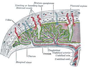
The feto-placental circulation continues to develop structurally and mature functionally throughout gestation as a result of continuing growth and maturation of villous trophoblast 9, whereas the structural remodeling of the utero-placental vasculature is considered to be complete shortly after 20-22 weeks of human pregnancy. Development of the placental vasculature. (A) Placental villi of 6 weeks gestational age prior to onset of the chorionic circulation, showing the presence of nucleated erythrocytes in the developing fetal capillaries (arrowed).(B) Villi at 14 weeks gestational age showing the presence of non-nucleated erythrocytes in the larger vessels within the stromal core, indicative of onset of the Structure of the placenta. There are certain unique features in the structure of the placenta and blood circulation in this organ that are directly relevant to pharmacologic actions of cocaine and amphetamines (Osol and Mandala, 2009).The placenta is of fetal origin and its blood circulation on the maternal side is established by destruction of blood vessels in the uterine tissue at the site
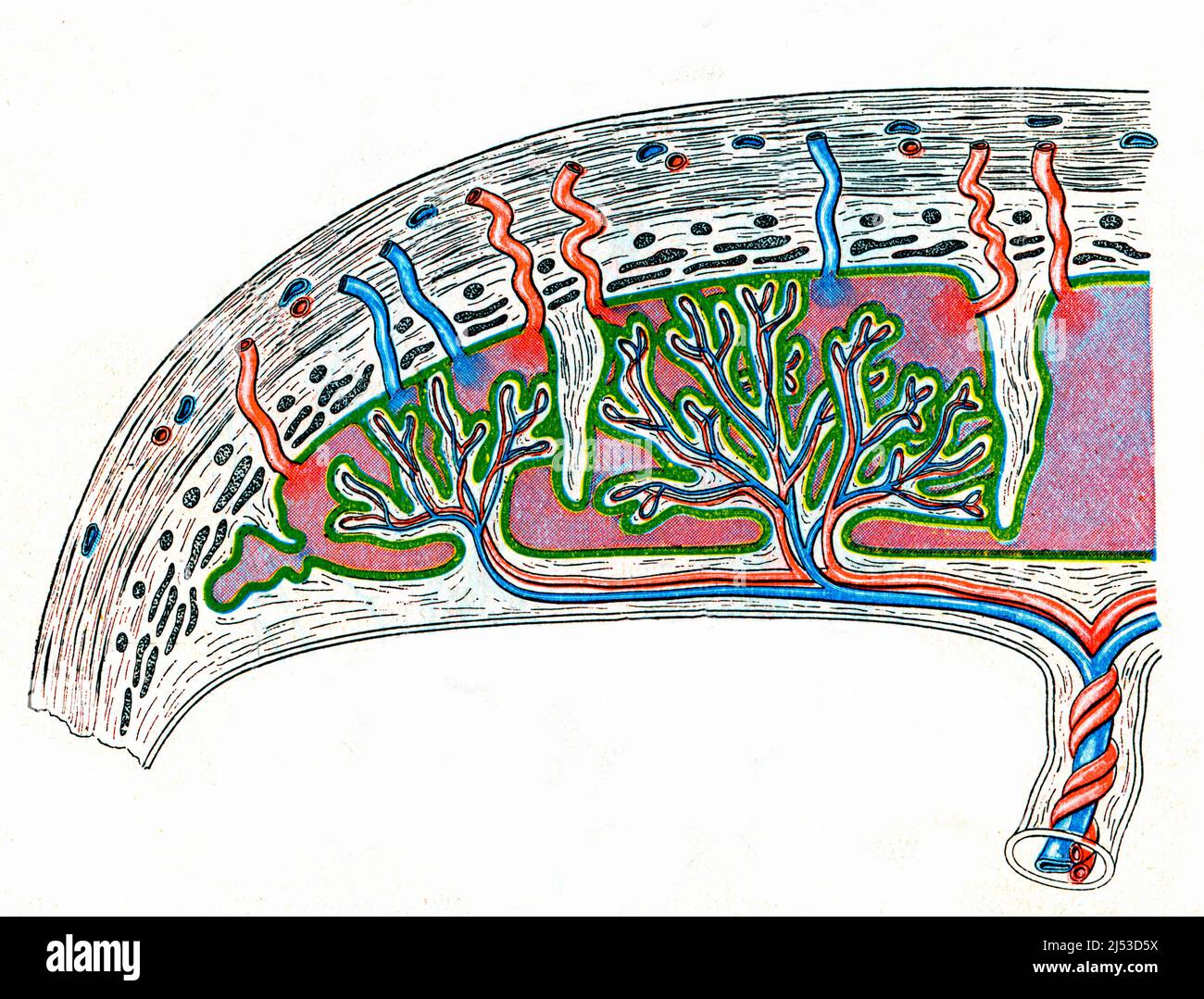
Placental blood circulation Biology Diagrams
The fetal circulation system is distinctly different from adult circulation. This intricate system allows the fetus to receive oxygenated blood and nutrients from the placenta. It is comprised of the blood vessels in the placenta and the umbilical cord, which contains two umbilical arteries and one umbilical vein. Fetal circulation bypasses the lungs via a shunt known as the ductus arteriosus

The placenta is the medium through which material passes from the maternal circulation to the fetal circulation by passive diffusion or active transport.[1] The placental growth factor is released from the placenta to prepare the mother's body for pregnancy in terms of cardiovascular adaption. (HCS), also known as human placental Most of the circulation to the lower body is supplied by blood passing through the ductus arteriosus. This blood then enters the umbilical arteries and flows into the placenta. In the placenta, carbon dioxide and waste products are released into the mother's circulatory system, and oxygen and nutrients from the mother's blood are released into There is a small body of literature suggesting that cyclooxygenase-2 (PTGS2) may play a role, although the data are conflicting as to whether it is upregulation or downregulation of PTGS2 that is resulting in abnormal vasoconstriction. Autacoids and the control of vascular tone in the human umbilical-placental circulation. Placenta. 1991;12
