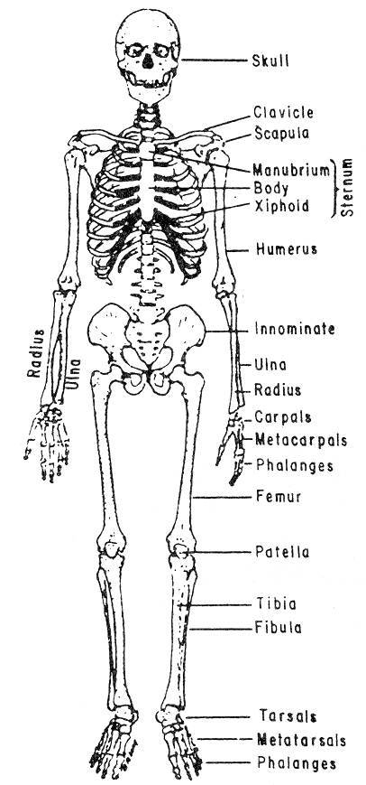
Naval Education and Training Command Hospital Corpsman 3 2 Chapter Biology Diagrams The human skeletal system is made up of more than 200 bones and has two main parts: the axial and appendicular skeleton. Find labeled diagrams here. Biopsy: For the skeletal system, this procedure can involve removing a small sample of bone or bone marrow so that it can be tested for conditions such as cancer. The skeletal system is your body's support structure. Its parts include your bones, muscles, cartilage and connective tissue like ligaments and tendons. The human skeleton is like a built-in suit of armor for organs throughout your body. Your skull protects your brain, human skeleton, Internal skeleton that serves as a framework for the human body.It consists of many individual bones and cartilages; there also are bands of fibrous connective tissue (the ligaments and tendons) in intimate relationship with the parts of the skeleton. The human skeleton has two main subdivisions: the axial, comprising the vertebral column and much of the skull, and the

The human skeleton is the internal framework of the human body.It is composed of around 270 bones at birth - this total decreases to around 206 bones by adulthood after some bones get fused together. [1] The bone mass in the skeleton makes up about 14% of the total body weight (ca. 10-11 kg for an average person) and reaches maximum mass between the ages of 25 and 30. [2] The Intricate Orchestra of Bones: The Human Skeletal System. Our bodies are a symphony of interconnected parts, and the skeletal system - a network of bones, is the conductor that orchestrates our every move. From the delicate fingers that gracefully manipulate objects to the sturdy legs that propel us forward, the skeleton is the framework

Skeletal System: What It Is, Function, Care & Anatomy Biology Diagrams
human skeleton, the internal skeleton that serves as a framework for the body. This framework consists of many individual bones and cartilages.There also are bands of fibrous connective tissue—the ligaments and the tendons—in intimate relationship with the parts of the skeleton. This article is concerned primarily with the gross structure and the function of the skeleton of the normal

Learn about the bones, joints, and skeletal anatomy of the human body with our interactive 3D models. Explore the axial and appendicular skeleton, the skull, the vertebrae, the ribs, the pectoral and pelvic girdles, and the upper and lower limbs. The human skeletal system consists of two primary divisions: the axial skeleton and the appendicular skeleton. Axial skeleton: The axial skeleton is composed of the skull, vertebral column, and rib cage.It offers a safeguard and shelter for the crucial organs of the body. The skull serves to protect the brain and provide support to the face. Learn about the skeletal system, which provides support, protection, movement and blood cell production for the body. Find out the anatomy and functions of bones, joints, cartilages and bone marrow in humans and other animals.
