Myocardium Endocardium And Pericardium Biology Diagrams This article will discuss the layers of the heart (the epicardium, the myocardium and the endocardium) and any clinical relations pertaining to them.. In the same way that vehicles have their fuel pumps, our body has the heart. The heart is a muscular organ found in the middle mediastinum that pumps blood throughout the body. It is housed in the pericardial sac, which protects it and assists Stocktrek Images/Getty Images. Epicardium (epi-cardium) is the outer layer of the heart wall.It is also known as visceral pericardium as it forms the inner layer of the pericardium. The epicardium is composed primarily of loose connective tissue, including elastic fibers and adipose tissue.The epicardium functions to protect the inner heart layers and also assists in the production of
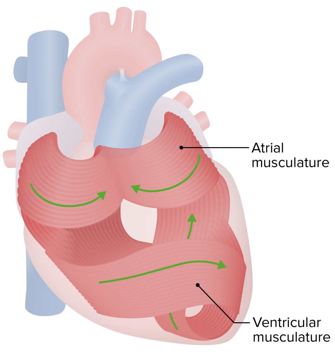
The main difference between myocardium and pericardium is that the myocardium is the muscular layer of the heart made up of heart muscles whereas the pericardium is the fibrous encase of the heart made up of connective tissue. Furthermore, the myocardium is responsible for heart contractions while pericardium is responsible for the protection of the heart. The myocardium and pericardium are two essential components of the heart, each with distinct attributes and functions. The myocardium, composed of specialized cardiac muscle tissue, is responsible for the heart's contractility, houses the electrical conduction system, and contains a rich vascular network.
The 3 Layers of the Heart Wall Biology Diagrams
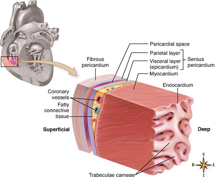
The parietal pericardium is the outer layer consisting of dense connective tissue, protecting the heart and upholding its position within the thoracic myocardium, and endocardium—are depicted, highlighting the structural relationships among them. This page titled 9.4: The Pericardium is shared under a CC BY 4.0 license and was authored Cardiac muscle (or myocardium) makes up the thick middle layer of the heart. It is one of three types of muscle in the body, along with skeletal and smooth muscle. The myocardium is surrounded by a thin outer layer called the epicardium (AKA visceral pericardium) and an inner endocardium. Coronary arteries supply to the cardiac muscle, and cardiac veins drain this blood. Cardiomyocytes are the The myocardium is the thickest of the three layers, particularly within the left ventricle, and is responsible for the ejection of blood during contractions. The epicardium (visceral pericardium) is responsible for the production of pericardial fluid and the protection of the other heart layers. Pericardium anatomy, Right Subclavian Artery
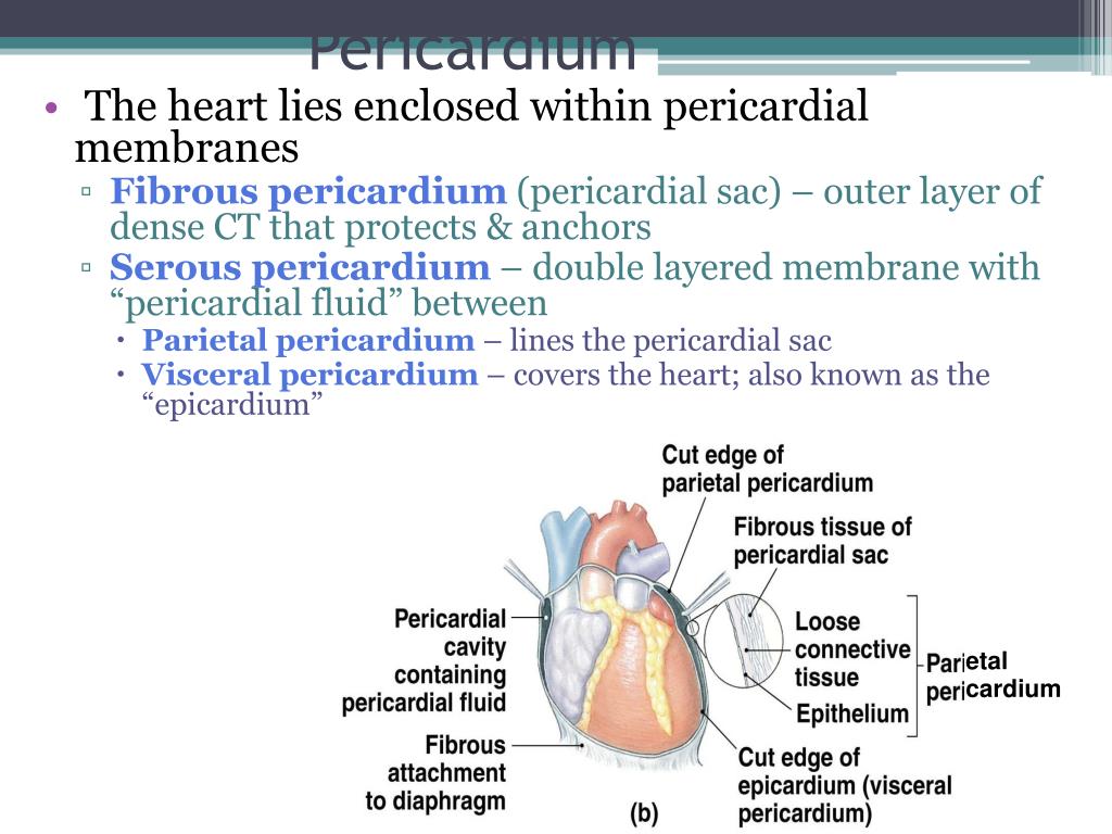
Location: Anatomically, it is located just beneath the parietal serous pericardium layer of the pericardium and is in direct contact with the myocardium. Epicardium Structure Histologically, it contains an outer layer of epithelium cells called the mesothelium below which is the subserosal layer made of connective tissues and a layer of fatty The myocardium and pericardium are both part of the heart wall, but they have distinct structures and functions. Myocardium vs. Pericardium: Key Differences Published in Heart Anatomy 2 mins read Feb 18, 2025 . The myocardium and pericardium are both part of the heart wall, but they have distinct structures and functions. Pericardium. The pericardium is a protective sac made of tough, flexible tissue that surrounds the heart. the epicardium, myocardium, and endocardium. These layers are similar to the three layers found in blood vessels, which are called the tunica adventitia, tunica media, and tunica intima. Knee Anatomy: Complete Guide to Parts, Names
