Hippocampus anatomy histology Biology Diagrams Hippocampus in the human brain Nissl-stained coronal section of the brain of a macaque monkey, showing hippocampal formation and subfields (circled). Hippocampus anatomy describes the physical aspects and properties of the hippocampus, a neural structure in the medial temporal lobe of each cerebral hemisphere of the brain.It has a distinctive, curved shape that has been likened to the sea
In summary, the third edition offers a slight improvement on the second edition; Duvernoy's second edition has been the gold standard for describing the anatomy of the human hippocampus. There is no other atlas that has such beautifully photographed images with corresponding diagrams and MR images as The Human Hippocampus. For those
Hippocampus: What It Is, Function, Location & Damage Biology Diagrams
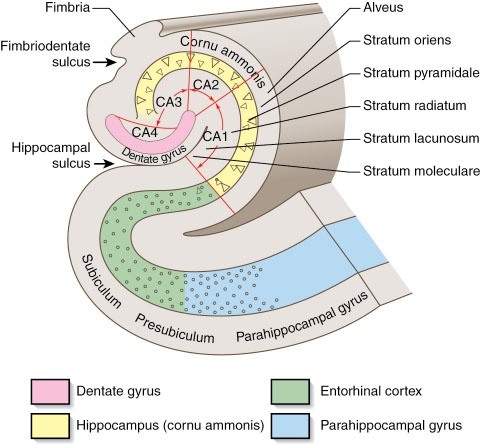
Anatomy of the hippocampus. The anatomy of the hippocampus is of chief importance to its function. The hippocampus receives input from and sends output to the rest of the brain via a structure known as the entorhinal cortex, which is located beneath the anterior (frontal) region of the hippocampus. The hippocampal formation itself is composed
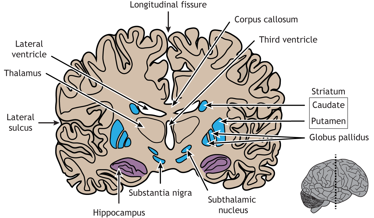
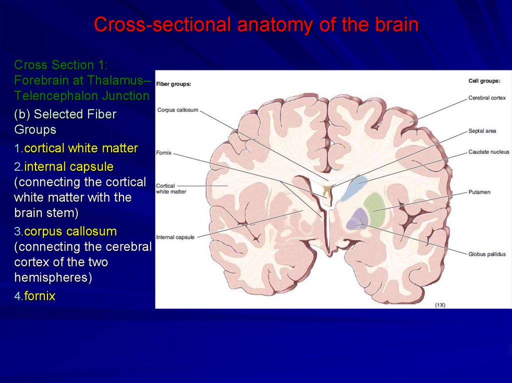
The hippocampus (pl.: hippocampi; via Latin from Greek ἱππόκαμπος, 'seahorse'), also hippocampus proper, is a major component of the brain of humans and many other vertebrates.In the human brain the hippocampus, the dentate gyrus, and the subiculum are components of the hippocampal formation located in the limbic system.The hippocampus plays important roles in the consolidation of Gross anatomy Location. The hippocampus lies in the hippocampal sulcus immediately below the floor of the temporal horn of the lateral ventricle, and in cross section (coronal) has appearances that are reminiscent of a seahorse.It has a head (posterior to the amygdala), a body, and a tail (which follows the upwardly curving lateral ventricle).. Although there is a lack of consensus relating to

Radiology Reference Article - Radiopaedia.org Biology Diagrams
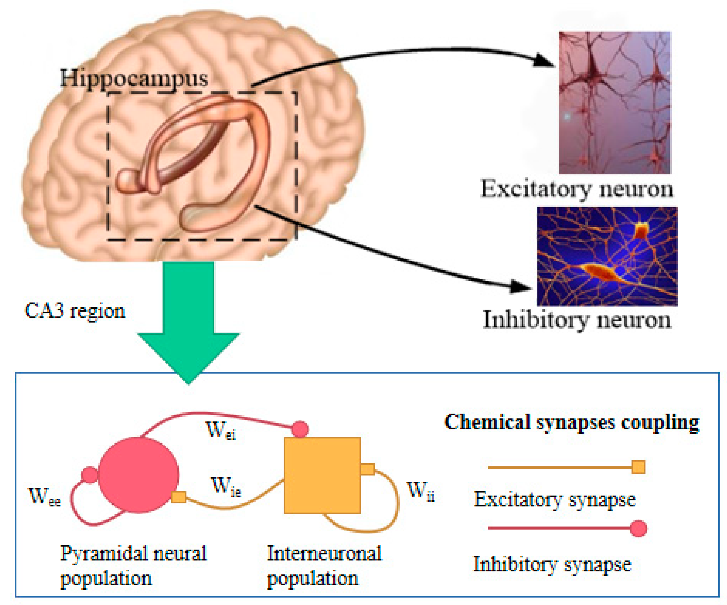
The hippocampal formation is responsible for memory processing, learning, spatial navigation, and emotions. It includes the indusium griseum, longitudinal striae, gyrus fasciolaris, hippocampus proper (cornu ammonis, dentate gyrus, and subiculum) and part of the uncus. The hippocampus has the archipallial cortex and is formed by the infoldings of the dentate gyrus, cornu ammonis and subiculum The hippocampus is the "flash drive" of the human brain and is often associated with memory consolidation and decision-making, but it is far more complex in structure and function than a flash drive. The hippocampus is a convex elevation of gray matter tissue within the parahippocampal gyrus inside the inferior temporal horn of the lateral ventricle. One can describe it more holistically as a This new edition, like previous ones, offers a precise description of the anatomy of the human hippocampus based upon neurosurgical progress and the wealth of medical imaging methods available. The first part describes the fine structures of the hippocampus and is illustrated with new original figures. A survey is then provided of current
The hippocampus is a paired structure present in each temporal lobe of the brain. It takes its name from the Greek word for the seahorse, because it resembles this small upright-swimming fish.. The hippocampus is part of a larger structure of the temporal lobe called the hippocampal formation.The hippocampal formation extends from the amygdala anteriorly, to the splenium of the corpus callosum Hippocampus proper (cornu ammonis): This region works to form memories, then organizes and stores your memories. Four regions help the hippocampus properly do its job, including cornu ammonis (CA) 1, 2, 3 and 4. Subiculum: This area collects information from the hippocampus to send messages to other parts of your brain to help with memory