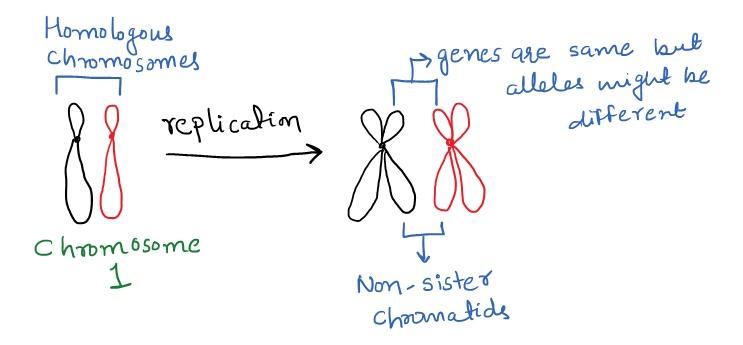
Chapter 5 The Cell Cycle and Meisois Biology Diagrams The paternal (blue) chromosome and the maternal (pink) chromosome are homologous chromosomes.Following chromosomal DNA replication, the blue chromosome is composed of two identical sister chromatids and the pink chromosome is composed of two identical sister chromatids.In mitosis, the sister chromatids separate into the daughter cells, but are now referred to as chromosomes (rather than While sister chromatids are exact copies of each other, non-sister chromatids come from homologous chromosomes. They code for the same genes, but are not genetically identical. domain contains a dynamic arrangement of proteins that are involved in mitotic checkpoints and regulators of chromosome behavior.

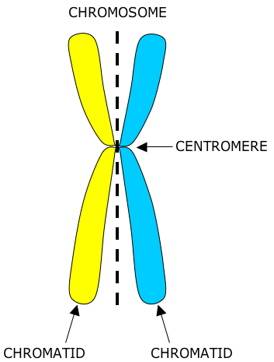
Centromere behavior also differs. In Anaphase I, cohesin proteins keep sister chromatids together, protected by Shugoshin. In Anaphase II, this protection is lost, allowing separase to cleave cohesin. The centromeres split, enabling chromatids to move independently. Errors and Resulting Consequences Sister chromatids, identical genetic copies of a chromosome, exhibit a fundamental behavior during cell division. These paired structures remain physically attached until a specific point in mitosis, known as anaphase. This separation is orchestrated by the spindle fibers, which are composed of microtubules that attach to structures called kinetochores on the centromeres of the chromosomes. Understanding the behavior of sister chromatids in mitosis is essential for grasping how cells divide and reproduce. Prophase. In prophase, the chromosomes condense and become visible under a microscope. Each chromosome appears as two sister chromatids joined at the centromere. The nuclear membrane starts to break down, and the mitotic spindle
Dynamics of sister chromatids through the cell cycle: Together and ... Biology Diagrams
Takahashi and Hirota preview work from Stanyte et al. examining the behavior of sister chromatids and their resolution as the cell cycle progresses. Dynamic organization of sister chromatids after replication. Replicated chromatin fibers are highly mobile and readily dissociate from each other for >300 nm, beyond the achievable resolution Advanced techniques allow researchers to observe sister chromatids' structure and behavior. Fluorescence microscopy uses dyes to visualize chromatids in live cells, while G-banding provides detailed chromosomal views. Confocal and super-resolution microscopy enhance visualization, and CRISPR-Cas9 tagging allows precise chromatid observation. The identical copies of each chromosome are known as sister chromatids, and they are tightly associated together through G2 phase and early mitosis. During metaphase of mitosis, sister chromatids are associated with the mitotic spindle, aligned along the central axis of the cell, and one sister from each pair is associated with a separate