75 best images about MRI Technology on Pinterest Biology Diagrams Weir and Abrahams' Imaging Atlas of Human Anatomy (Includes Videos) by Jonathan Spratt; Lonie R. Salkowski; Jamie Weir; The perfect up-to-date imaging guide for a complete and 3-dimensional understanding of applied human anatomy Imaging is ever more integral to anatomy education and throughout modern medicine. Building on the success of previous editions, this fully revised sixth edition Weir and Abrahams' Imaging Atlas of Human Anatomy, 6th edition by Jonathan D. Spratt; Lonie R. Salkowski; Marios Loukas; Tom Turmezei; Langman's Medical Embryology, 15th edition by T. W. Sadler. ISBN: 9781975179960. Publication Date: 2024. UTMB Links. Gray's Anatomy for Students, 5th edition by Richard L. Drake (Editor); A. Wayne Vogl All lessons are designed to emphasize important imaging anatomy for health professionals with particular focus given to cross-sectional anatomy and variants (course trailer, lecture topics). Ideal for all health professionals and students, particularly those approaching an examination in anatomy. Key Features. 8 hours of expert video teaching

• Uses concise, brief text that explains the condition, thus allowing the radiologic images to guide you to the differentiating factors. • Incorporates discussions of imaging modality choices for a range of pathologies to help you understand how to select imaging procedures for various clinical situations in the clinical environment. Diagnostic Imaging principles and concepts are augmented by the presentation of images for common clinical conditions. Guiding principles related to minimizing radiation exposure and requesting the most appropriate imaging examination are addressed.Static images are enhanced by the ability to access images stored and displayed on an Html-5 compatible, Dicom image viewer that simulates a simple

Anatomy, the Anatomy of Imaging Biology Diagrams
Designed for busy medical students, The Radiology Handbook is a quick and easy reference for any practitioner who needs information on ordering or interpreting images.The book is divided into three parts:- Part I presents a table, organized from head to toe, with recommended imaging tests for common clinical conditions.- Part II is organized in a question and answer format that covers the Radiology Masterclass - Provides an overview of medical imaging, complete with tutorials and access to images. Targeted at both medical students and other health professionals, this is currently a free UK resource. Topics include x-ray physics, anatomy visible in x-rays, and trauma x-rays. e-Anatomy is a high-quality anatomy and imaging content atlas.It is the most complete reference of human anatomy available on the Web, iPad, iPhone and Android devices. Explore detailed anatomical views and multiple modalities (over 8,900 anatomic structures and more than 870,000 translated medical labels) with images in CT, MRI, radiographs, anatomical diagrams and nuclear images.
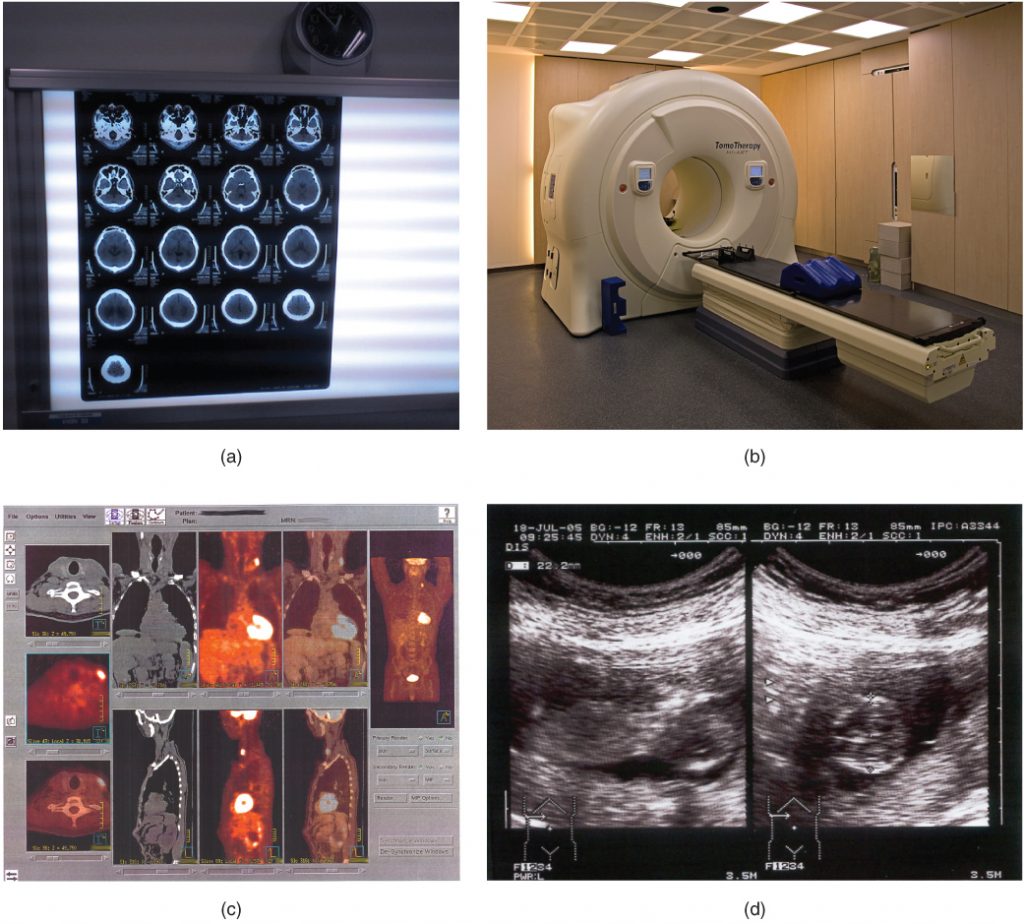
Keywords: X-ray; Interpretation; Anatomy; Chest; Fracture; Imaging. The Science Behind X-Rays. X-rays were discovered in the late 19th century by Wilhelm Conrad Röntgen. Their unique ability to pass through certain materials—such as soft tissue—while absorbed by denser tissues, such as bone, makes them especially useful in medical imaging. Normal chest x ray. Radiological anatomy is where your human anatomy knowledge meets clinical practice. It gathers several non-invasive methods for visualizing the inner body structures. The most frequently used imaging modalities are radiography (X-ray), computed tomography (CT) and magnetic resonance imaging (MRI).X-ray and CT require the use of ionizing radiation while MRI uses a magnetic
